5.03 Red legs, Swollen legs
5.03 – Red Legs, Swollen Legs
Presented by: Dr Jo Preston, Dr Iain Wilkinson with Dr Bernard Ho and Dr Jonathon Slater
Broadcast Date: 6th March 2018
Click here for a pdf of the sip of MDTea poster
Click here for a pdf of show notes
Social Media this week
Guess what happens in 2 months?
It is World Delirium Awareness Day! 14 Mar 2018
Awake, Aware, Action – Plan your events now! Please tag #WDAD2018 & @iDelirium_Aware pic.twitter.com/EoRlbbopBV
— iDelirium (@iDelirium_Aware) January 14, 2018
In ‘The Gallery’ this week
Etching by Freya Payne – if can be seen here
To see more of her works have a look at the flowers gallery here
ADDITIONAL RESOURCES
CPD log
Click here to log your CPD online and receive a copy by email.
Tweetchat #MDTeaClub
We will be hosting a ‘journal club’ type tweet chat to discuss topics raised in this episode using #MDTeaClub
Join us to discuss topics raised in the episode and spread any resources you may have!
Main Show Notes:
Learning Outcomes
Knowledge:
- To recall the common causes of red legs and how they are managed
- To recall the common causes of swollen legs
- To know the causes of lymphoedema
Skills:
- To recognise when red legs are not cellulitis
- To identify lymphoedema
Attitudes:
- To appreciate that accurate diagnosis of both red legs and swollen legs is important
- To see these presentations as opportunities to provide advice on preventative care to prevent complications of minimise recurrence.
Key Points from Discussion
Red Legs
Cellulitis
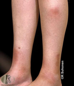
Cellulitis is an infection of the skin so will be associated with features of infection such as fevers and potentially sepsis too. The onset will be rapid onset of unilateral, progressive redness.
Predisposing factors
- Presence of lymphoedema
- Previous cellulitis
- Diabetes
- Immunosuppression
Often caused by a pathogen on the skin gaining entry beneath, so look for and ask about skin breaks including insect bites and fungal infections and check between the toes. Ensure good foot care and maintaining good skin care including adequate moisture barrier maintained to prevent further infections.
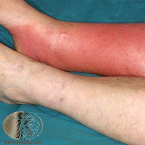
Image courtesy of dermnetnz.org
Treatment is with antibiotics. If more than two episodes in a year then can consider prophylaxis.
Penicillin to Prevent Recurrent Leg Cellulitis
Antibiotic prophylaxis for preventing recurrent cellulitis: a systematic review and meta-analysis. Oh et. al, Journal of Infection 2014
Lipodermatosclerosis
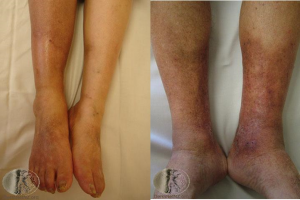
An inflammatory condition of the lower legs usually due to venous insufficiency. It is usually a deep red colour, compared to the bright pink of a cellulitis. Acute flares can happen and can be red, painful and scaly. Will not be associated with raised inflammatory markers and does not have an acute progression. It is usually bilateral, which cellulitis rarely is.
Chronic lipodermatosclerosis is associated with increased swelling in the leg, moderate redness, increased pigmentation and atrophe blanche (small white areas).
Below are pictures of acute (left) and chronic (right) lipodermatosclerosis, courtesy of dertnetnz.org
Pannicullitis
An inflammation of the subcutaenous fat, so a much deeper inflammatory process. They are painful, and often ill defined.
Often a presenting feature a systemic problem, rather than one locally in the leg. Can be seen in many conditions including Inflammatory Bowel Disease, sarcoidosis and some drugs can induce it, such as NSAIDS and the oral contraceptive pill.
Image courtesy of dermnetnz.org
Swollen Legs
Fluid Overload
This is commonly seen as a consequence of heart failure. If this happens quite quickly it can often appear red and may blister. Usually though it will just be a swelling of the skin with subcutaenous fluid. It will be bilateral and pitting. The history will usually help to differentiate this. Treatment of the underlying cause, so in this case, through diuresis to remove the excess fluid as well as good wound care of any breaks to prevent deterioration.
Dependent Oedema
It can be difficult to determine the difference between fluid overload and dependent oedema. In dependent oedema, the fluid tends to predominantly follow gravity, so if the legs are flat on the bed, the undersides will be swollen and pitting, and this will change to be feet and ankles on sitting in a chair. It tends not to rise up the legs so far as fluid overload can. Investigations for heart failure including blood tests such as BNP and an echocardiogram can help to rule out heart failure which requires diuretic therapy.
The mainstay of advice is usually to elevate the legs closer to the level of the heart to help the fluid to be cleared from the legs. There is some debate about this as a treatment….
Interview on the debate – dermatologist at ESH?
Low Protein States: Malnutrition, Critical Illness and Nephrotic Syndrome.
Low protein can be seen in severe malnutrition and when a person has been critically unwell, usually with a severe illness or prolonged physiological stressor. Examples of when this is commonly seen include after an ITU stay or emergency surgery with long recoveries. Dietician involvement is important to aid recovery.
Low protein states cause oedema in all extremities, including the upper limbs but will also, within this, be worse in dependent areas. Other causes are not usually seen in the upper limbs.
Nephrotic syndrome is a particular condition in which the kidneys release excess protein leading to a whole body depletion of stores. This can be picked up on a urine dipstick for protein which will be highly positive. In nephrotic syndrome, the pattern of oedema will often include the arms and sometimes the face.
Drugs
Drugs which cause fluid retention, for example blood pressure medications such as amlodipine (a calcium channel blocker), can result in swollen legs.
Lymphoedema
Lymphoedema is a condition affecting the lymphatic system of the body, a network of channels and glands which help fight infection and remove excess fluid. It can be primary (due to a problem in the development of the lymphatic system (presents in early adulthood at latest) or secondary.
Secondary causes are due to damage to the lymphatic system.
- Following lymph node surgery or radiotherapy treatment of cancers
- Severe cellulitis can cause scarring of the lymphatic system
- Inflammatory conditions such as RA or eczema for same reason
- Venous disease (including DVT and varicose veins) – abnormal or damaged veins causes overflow of fluid from veins into tissue spaces which overwhelms and eventually exhausts the lymphatic system involved.
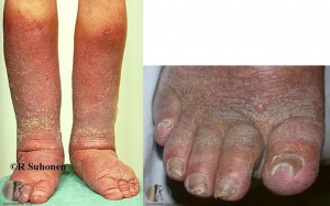
In its early stages, lymphoedema can be pitting and resemble fluid overload. In the latter stages skin becomes hardened and tight resulting in deep, chronic lines. In particular ‘squaring of the toes’ can be an early sign of this.
Images courtesy of dermnetnz.org
Mainstay of treatment is compression bandaging. This requires the exclusion of significant arterial insufficiency before applying, through either a doppler or an ABPI, depending on which is available.
Lipoedema
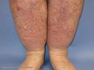
This is an abnormal accumulation of adipose tissue (fat) predominantly in the lower limbs which is gaining recognition. It fails to respond to compression and may initially be mistaken for lymphoedema.
Image courtesy of dermnetnz.org
Lipoedema UK: http://www.lipoedema.co.uk/
RCGP e-learning course on Lipoedema
Curriculum Mapping:
This episode covers the following areas (n.b not all areas are covered in detail in this single episode):
| Curriculum | Area | |
| NHS Knowledge Skills Framework | Suitable to support staff at the following levels:
| |
| Foundation curriculum | Section
2.1 2.2 3.10 3.16 | Title
Patient as centre of care Communication with patients Recognises, assesses and manages patients with long term conditions Demonstrate understanding of health promotion and illness prevention |
| Core Medical Training | Dermatology
Limb Pain & Swelling Managing long term conditions | |
| GPVTS program | Section 2.03 The GP in the Wider Professional Environment
Section 3.05 – Managing older adults
| |
| ANP (Draws from KSF) | Section 7.31 – Problems with skin | |
| Geriatric Medicine Training Curriculum | 29. Diagnosis and Management of Chronic Disease and Disability
38. Tissue Viability |
